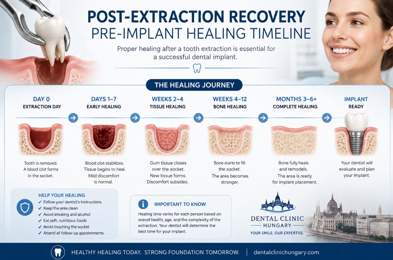
What to Expect After Tooth Extraction: Quick Summary
After a tooth extraction, the socket first forms a protective blood clot, then develops pale granulation tissue and gradually closes with gum. Although the gum may look healed within a few weeks, the jawbone underneath continues to remodel for three to six months.
-
Normal healing: Pain and swelling gradually improve. White, cream or pale-yellow tissue is often normal granulation tissue, not pus.
-
Dry socket: Usually causes severe, worsening pain a few days after extraction when the blood clot is lost too early. It often needs dental cleaning and a soothing dressing.
-
Infection: More likely if pain and swelling worsen, especially with pus, fever, facial swelling, foul taste or feeling unwell. Contact a dentist promptly.
-
Ridge preservation: A bone graft placed immediately after extraction can reduce jawbone shrinkage and may make a future implant easier and more predictable.
-
Implant timing: An implant may be placed on the same day, after several weeks, or after three to six months of healing. The best option depends on infection, bone quality, gum condition and implant stability.
-
Trismus: Jaw stiffness after surgery is common, particularly after difficult lower-tooth extractions. It usually settles as swelling reduces, but worsening stiffness or difficulty swallowing requires urgent dental advice.
The key message: protect the healing socket, do not disturb pale healing tissue, and seek professional advice if symptoms worsen rather than improve.
Post-Extraction Recovery and Pre-Implant Healing in details
A tooth extraction is not the end of treatment – it is often the beginning of a carefully planned healing period. Whether you are considering a future dental implant, have had a socket graft, or are simply worried about how the extraction site looks and feels, understanding the first days, weeks and months after removal can make recovery less stressful.
The most important point is this: the gum may look healed long before the jawbone has fully healed. During the first three to six months after extraction, the socket changes significantly. Bone remodels, gum tissue matures and the ridge may shrink in height and width. This is why dentists may recommend ridge preservation at the extraction appointment, delayed implant placement after healing, or—in selected cases – an immediate implant.
This UK-focused guide explains normal socket healing, bone preservation, dry socket and infection warning signs, implant timing, and trismus (jaw stiffness) after extraction or implant surgery.
Important: This article is general information, not a diagnosis. Contact your dentist, oral surgeon or implant clinician for advice tailored to your procedure and medical history.
The First Days After Extraction
The empty space left after a tooth is removed is called an extraction socket. It is a wound in both the gum and the tooth-bearing jawbone, also known as alveolar bone.
Healing begins immediately.
First 24 Hours: Blood Clot Protection
A blood clot forms inside the socket after extraction. This clot is the body’s natural dressing: it controls bleeding, covers sensitive bone and nerve endings, and provides the foundation for new healing tissue.
The clot may appear dark red, brown-red or almost black. This can be normal. The most important aftercare goal is protecting it during the first day.
General advice usually includes:
-
Bite on gauze for as long as your dental team instructs.
-
Rest after treatment and avoid strenuous exercise.
-
Do not rinse your mouth or spit forcefully for 24 hours.
-
Avoid smoking, vaping and alcohol.
-
Do not drink through a straw.
-
Do not touch the socket with fingers, toothbrush bristles or your tongue.
-
Eat soft, cool or lukewarm foods once the numbness has worn off.
These measures reduce the chance of disturbing the clot. A lost or prematurely broken-down clot can lead to dry socket, a painful post-extraction complication.
Days 2 to 3: Pain and Swelling May Peak
Mild to moderate pain, tenderness, swelling and limited mouth opening can be expected after an extraction, especially after impacted wisdom tooth removal, surgical extraction, socket grafting or implant surgery.
For many patients, swelling is most noticeable at around 48 to 72 hours before gradually settling. This means that looking slightly more swollen on day two than on the evening of surgery is not automatically a sign of infection. The overall pattern is more important than any single day: discomfort should stabilise and gradually improve rather than continuously intensify.
Use the pain relief advised by your dentist, pharmacist or GP. Paracetamol and ibuprofen are frequently used after oral surgery, but ibuprofen is not suitable for everyone. Ask a healthcare professional if you have a history of stomach ulcers, kidney disease, asthma affected by anti-inflammatory medicines, bleeding problems, heart conditions or medication interactions.
Days 4 to 7: Granulation Tissue Develops
By around days four to five, the original blood clot begins to be replaced by granulation tissue. This is healthy repair tissue containing small blood vessels, collagen and healing cells.
Granulation tissue may look:
-
White, off-white or cream-coloured
-
Pale yellow or yellowish-white
-
Pink, red or mottled
-
Soft, slightly uneven or lumpy
This appearance often causes unnecessary anxiety. A pale or yellowish layer is not automatically pus. It may be fibrin, a protein involved in normal wound repair, or granulation tissue covering the healing socket.
Do not scrape, pick or rinse aggressively to remove it. Interfering with healthy healing tissue can cause bleeding, pain or delayed recovery.
Weeks 1 to 2: Gum Healing
During the first one to two weeks, the gum edges gradually move over the socket. The hole may still be visible, particularly after a molar or wisdom tooth extraction, and food may occasionally collect there.
From the day after extraction, gentle warm salt-water rinses are often advised, although your own clinician’s instructions always come first. Brush the rest of the mouth normally and clean carefully around the surgical area. Avoid driving toothbrush bristles deeply into the socket.
Surface gum closure may be well under way after a few weeks, but the bone beneath it continues to heal and remodel for months.
What Happens to Jawbone in the First 3 to 6 Months?
The bone around a tooth exists partly because the tooth root is there. Once the root is removed, the supporting alveolar ridge begins to remodel naturally.
This process is normal. It does not mean that an extraction has “gone wrong”. However, it matters if you are considering an implant, fixed bridge or removable denture later.
Why the Ridge Shrinks
The extraction socket fills with blood clot, then granulation tissue, immature bone and eventually more mature bone. At the same time, the outer walls of the ridge can shrink—especially the thin bone on the cheek side of the socket.
This cheek-side, or facial, bone plate is often naturally thin. After a tooth is removed, it can resorb more than the palatal or tongue-side bone. The gum follows the bone contour, so bone loss can also flatten the surrounding gum profile.
Research has found substantial dimensional changes in untreated extraction sites. A systematic review reported horizontal bone reduction of approximately 32% by three months and 29% to 63% by six to seven months; vertical loss at six months was reported at around 11% to 22%.
The Three-Month Point
By approximately three months, the socket may look completely closed at the gum surface, yet deeper bone remodelling is still continuing. This is often when an implant clinician reassesses the area using an examination, X-rays or a CBCT scan.
For some people, there is enough bone for implant placement at this stage. For others, especially where the original tooth had infection, gum disease, a thin facial bone wall or a difficult extraction, a longer healing period or additional grafting may be better.
The Six-Month Point
At around four to six months, bone healing is more mature and implant planning can be more predictable. The clinician can assess the ridge’s height, width and position, as well as the gum contour needed for a functional, natural-looking implant crown.
Bone healing is not identical for everyone. The upper and lower jaw heal differently, graft materials have different remodelling characteristics, and health factors such as smoking, diabetes and gum disease can influence the process.
Socket Preservation: Why Immediate Bone Grafting May Help
Socket preservation, also called ridge preservation or socket grafting, is a preventive procedure performed when a tooth is removed. It aims to reduce post-extraction collapse of the jaw ridge.
After a tooth is extracted, the clinician may place bone graft particles into the fresh socket. A collagen membrane, collagen plug or gum-tissue covering may protect the graft, and stitches may be used to secure the area.
The goal is not to guarantee that the jawbone will never change. No graft can completely stop normal remodelling. Instead, the aim is to preserve more height, width and contour than would be expected if the socket were left to heal unaided.
Socket preservation is especially worth discussing when:
-
You want a dental implant later.
-
The tooth is at the front of the mouth and appearance is important.
-
The facial bone wall is thin, missing or damaged.
-
The extraction is expected to be difficult or surgical.
-
Gum disease or infection has already damaged the surrounding bone.
-
The implant will be delayed for medical, financial or planning reasons.
-
A bridge pontic or denture will need good ridge support.
The British Dental Journal notes that socket preservation helps maintain bone volume when implant placement must be delayed and may reduce the need for later, more extensive augmentation.
Graft Materials Explained
A socket graft may use:
Ask your dentist exactly what material is proposed. This is particularly relevant if you have religious, ethical or personal preferences about donor- or animal-derived products.
Does Every Extraction Need a Bone Graft?
No. Ridge preservation should be a clinical decision, not an automatic add-on.
It may be unnecessary if you do not intend to replace the tooth with an implant, if the ridge is likely to remain adequate for your chosen restoration, or if an immediate implant can be placed safely. It may also be unsuitable in some health or treatment circumstances.
However, if a future implant is likely, the best time to discuss bone preservation is before the tooth is extracted. Once bone has already resorbed, rebuilding it can involve a larger graft, more surgery, longer healing and higher cost.
Systematic-review evidence supports ridge preservation as a way to reduce post-extraction ridge-volume loss, although it does not eliminate all changes and the clinical result depends on the individual site and technique.
Normal Healing vs Dry Socket vs Infection
A healing socket can look unattractive. Colour alone is not a reliable way to diagnose a complication. Use the symptom pattern, timing and overall trend—and call your dentist if you are unsure.
Normal Granulation Tissue
Healthy granulation tissue is usually attached within the socket. It does not ooze like liquid, and it is not typically associated with fever, a rapidly enlarging swelling or severe worsening pain.
Do not assume that “white stuff” is infection. In many patients, it represents normal repair tissue.
Dry Socket
Dry socket, medically called alveolar osteitis, occurs when the protective clot is lost, dislodged or breaks down too early. The socket may appear empty, and the underlying bone can become exposed.
The most typical feature is severe, deep, persistent pain that develops or sharply worsens after a brief initial improvement. Pain can radiate towards the ear, temple, cheek or jaw on the same side. A bad taste or unpleasant smell may also occur.
Dry socket is painful but does not automatically mean infection. SDCEP guidance describes dry socket as inflammation after extraction and recommends measures such as suitable analgesia, saline irrigation and a dressing where required. It does not recommend routine antibiotics unless there are signs of spreading infection, systemic illness or a relevant clinical reason.
Infected Extraction Socket
An infection is more likely when worsening pain comes with increasing redness, warmth, facial swelling, genuine pus, fever, chills, swollen glands or feeling generally unwell.
Pus may be thick, creamy, yellow, yellow-green or white. Unlike attached granulation tissue, it may drain or ooze and is often accompanied by a persistent unpleasant taste or smell.
A dental infection needs assessment because treatment focuses on addressing the source. This can involve cleaning or irrigating the socket, draining pus, removing trapped debris or a retained fragment, and prescribing antibiotics only where clinically appropriate.
Antibiotics alone are not a substitute for local dental treatment. SDCEP advises local measures first for infections without systemic or spreading signs, with antibiotics used where infection is spreading, systemic symptoms are present, or the patient is immunocompromised.
Implant Placement: Immediate, Early or Delayed?
The phrase “same-day implant” can be confusing. It may refer to placing the implant post on the day of extraction, fitting a temporary tooth on the same day, or both. These are different steps.
Immediate Implant Placement
An immediate implant is placed in the fresh extraction socket at the same appointment as tooth removal. It can shorten the calendar timeline and may help maintain the gum and ridge contour, particularly in front-tooth cases.
However, immediate placement is not suitable for every patient. The clinician needs enough healthy bone to achieve strong initial stability, favourable socket anatomy, controlled infection, appropriate gum conditions and a bite that will not overload the implant.
An immediate implant may receive a temporary tooth straight away, but it may also be left unloaded beneath the gum. Immediate placement does not guarantee immediate loading or an immediate final crown.
Early Implant Placement
Early placement usually occurs after soft-tissue healing, often at around four to eight weeks, or after partial bone healing at roughly 12 to 16 weeks.
This can be useful when the dentist wants initial inflammation to settle while avoiding the full period of ridge remodelling associated with a completely delayed approach. It may be considered after extraction of a tooth with local infection, provided the site has healed favourably and there is adequate bone.
Delayed Implant Placement
Delayed placement often means waiting around three to six months after extraction. This is common when infection, poor bone volume, damaged socket walls, grafting needs or complex gum conditions make a staged approach safer.
A delayed implant is not an inferior implant. In fact, it can be the most predictable option in a compromised site because it allows time to rebuild bone, mature the gum and create a stable foundation for the implant.
Clinical reviews describe immediate, early and delayed protocols as viable options, with the decision based on case selection rather than patient pressure for the fastest route.
Osseointegration: Another Healing Stage
After the implant is inserted, it needs to integrate with the jawbone. This process is called osseointegration.
In straightforward cases, healing often takes around three to four months before the final crown is fitted. Grafted sites, sinus-lift cases, lower-density bone or patients with specific risk factors may require longer.
A realistic extraction-to-final-crown timeline can therefore range from around three to four months in a carefully selected immediate-placement case to six to 12 months or longer where grafting and delayed placement are needed.
Trismus: Lockjaw After Extraction or Implant Surgery
Trismus means restricted mouth opening due to jaw-muscle spasm, inflammation, swelling or discomfort around the jaw joint. Patients often describe it as “lockjaw”, although the jaw is not always truly locked.
It is relatively common after lower wisdom tooth surgery, lengthy surgical extractions and more extensive implant or bone-grafting procedures. Keeping the mouth open for a prolonged time, working near the chewing muscles and post-operative inflammation can all contribute.
Common Symptoms
-
Difficulty opening the mouth as wide as usual
-
Tightness in the cheek or jaw muscles
-
Pain when yawning, chewing or brushing back teeth
-
A feeling that the jaw is stiff or pulls to one side
-
Reduced ability to eat normal foods
-
Soreness near the jaw joint or temple
After lower third-molar surgery, some degree of pain, swelling and trismus is common, and studies report that reduced mouth opening often improves within seven to 10 days.
What Can Help?
Follow the instructions from your dental team. General measures may include:
-
Use prescribed or recommended pain relief appropriately.
-
Choose soft foods temporarily.
-
Apply a wrapped cold pack during the early inflammatory period if advised.
-
After the acute phase, use gentle warmth if muscle tightness remains and your clinician agrees.
-
Avoid forcing the jaw open.
-
Start gentle jaw movements or stretching only when your clinician recommends it.
-
Maintain careful oral hygiene without overstraining the jaw.
Evidence reviews note that pain relief, selected anti-inflammatory approaches and physical measures may help, but no one method reliably prevents all post-operative pain, swelling and trismus.
Do not attempt aggressive self-stretching, force the mouth open with objects or use unapproved muscle-relaxing products. If exercises are needed, a dentist, oral surgeon or physiotherapist can show you a safe programme.
When Trismus Needs Urgent Advice
Contact your clinic promptly if restricted opening is getting worse rather than better, persists beyond the recovery period expected for your procedure, or appears alongside infection symptoms such as increasing swelling, fever, pus, difficulty swallowing or feeling unwell.
Seek urgent help via your dentist, NHS 111 or emergency services if you have:
-
Difficulty breathing or swallowing
-
Rapidly spreading facial, jaw or neck swelling
-
A high temperature with worsening mouth opening
-
Severe pain that is not controlled by advised medication
-
A jaw that is stuck open or cannot be closed
-
Significant facial trauma
Protecting Your Healing Site
Good recovery is not about trying to make the socket look perfect each day. It is about creating the right conditions for healing and recognising when the trend is wrong.
During recovery:
-
Follow the aftercare sheet from your own dental provider.
-
Avoid smoking and vaping, particularly with nicotine.
-
Keep the mouth clean without disturbing the socket.
-
Attend review appointments, especially after grafting or implant surgery.
-
Use temporary teeth exactly as instructed.
-
Avoid biting hard foods directly on an implant or graft site.
-
Tell your dentist about worsening pain, swelling, discharge, fever or persistent jaw stiffness.
Final Takeaway
Post-extraction healing is a staged biological process. A dark clot, followed by white, cream, pale yellow or pink healing tissue, is often normal. The socket may close at the gum surface within weeks, while jawbone remodelling continues for three to six months and beyond.
Socket preservation can reduce the bone loss that commonly follows extraction and may make a future implant more straightforward. Immediate implants can work well in carefully selected cases, while early and delayed placement are often safer when infection, bone loss or grafting needs are present.
The best outcome comes from planning ahead: discuss your future tooth-replacement goals before extraction whenever possible, protect the healing socket afterwards, and contact your dentist promptly if recovery is worsening rather than improving.
Post-Extraction Recovery and Pre-Implant Healing FAQs
How long does it take for an extraction socket to heal?
The gum surface often begins to close within one to two weeks, but the bone underneath continues healing and remodelling for several months. If you are planning a dental implant, your clinician may reassess the site at around three to six months, depending on the extraction, bone condition and whether grafting was used.
What should a normal healing extraction socket look like?
A normal socket commonly progresses from a dark-red blood clot to white, cream, pale-yellow, pink or red granulation tissue. It may still look like a small hole for several weeks, particularly after molar or wisdom tooth removal. Appearance alone is not a reliable way to diagnose a problem; gradual improvement in pain and swelling is more reassuring.
Is white or yellow tissue in my extraction socket normal?
Often, yes. White, creamy or pale-yellow material may be fibrin or granulation tissue, both of which can be normal parts of healing. Do not attempt to scrape or remove it. Contact your dentist if it is associated with increasing pain, pus, swelling, fever or feeling unwell.
What is granulation tissue after tooth extraction?
Granulation tissue is newly forming healing tissue made up of tiny blood vessels, collagen and repair cells. It gradually replaces the original blood clot and helps the socket heal. It can appear pale, yellowish, pink or red.
How can I tell if my extraction socket is infected?
An infection is more likely if pain and swelling are getting worse rather than better, especially if you notice thick discharge or pus, persistent foul taste, increasing redness, fever, chills or feeling generally unwell. Arrange a prompt dental assessment rather than trying to treat or drain the site yourself.
What is the difference between dry socket and infection?
Dry socket usually causes severe, deep pain that begins or worsens a few days after extraction when the protective blood clot is lost too early. Infection is more likely to involve increasing swelling, redness, pus, fever or malaise. Both require dental advice, but they are different conditions and may be treated differently.
Can I get dry socket after a bone graft?
Yes, although careful surgical technique and following aftercare advice reduce the risk. Protect the blood clot and graft by avoiding smoking, vaping, straws, forceful spitting and vigorous rinsing during the early healing period.
Why does my jaw feel stiff after extraction or implant surgery?
Jaw stiffness is called trismus. It can develop because of swelling, inflammation, muscle spasm or keeping the mouth open during a lengthy procedure. It is particularly common after lower wisdom tooth surgery and usually improves as postoperative swelling settles.
How long does trismus last after dental surgery?
Mild jaw stiffness often improves within about seven to 10 days, although recovery can take longer after complex extractions, grafting or implant surgery. Contact your clinic if mouth opening is worsening, is not improving as expected or occurs with fever, increasing swelling or difficulty swallowing.
What is socket preservation or ridge preservation?
Socket preservation is a procedure performed at the time of extraction to reduce loss of jawbone volume. Bone graft material is placed into the socket, often protected by a collagen membrane or plug, to help maintain the ridge for a future implant, bridge or denture.
Do I need a bone graft after every tooth extraction?
No. A bone graft is not needed after every extraction. It may be recommended when you plan an implant later, where the socket walls are thin or damaged, where bone loss already exists, or where preserving gum and bone contour is especially important.
Why does jawbone shrink after extraction?
The bone around a tooth root, called alveolar bone, naturally remodels after the tooth is removed. The ridge can lose width and height, particularly in the first three to six months. Ridge-preservation grafting aims to reduce, but cannot completely prevent, this change.
Can I have a dental implant on the same day as extraction?
Sometimes. Immediate implant placement may be possible when there is adequate healthy bone, good initial implant stability, suitable gum conditions and no uncontrolled infection. It is not appropriate for every extraction, and a delayed plan may offer a more predictable result in complex cases.
How long after extraction can I have a dental implant?
An implant may be placed immediately, after several weeks of early healing, or after three to six months of more complete bone healing. If substantial grafting or infection treatment is needed, the timeline can be longer.
Will I need to wait after the implant is placed before getting a crown?
Usually, yes. The implant needs time to integrate with the jawbone, a process called osseointegration. In straightforward cases this often takes around three to four months, but grafted or complex sites may need longer.
What should I avoid after extraction, grafting or implant surgery?
Avoid smoking and vaping, alcohol, straws, forceful rinsing, forceful spitting and hard foods during the early recovery period. Follow your own dentist’s instructions, protect the surgical site and avoid chewing directly on a graft or implant while it heals.
When should I call my dentist urgently?
Contact your dentist promptly for worsening pain, increasing swelling, persistent bleeding, suspected pus, fever, foul taste with worsening symptoms, a loose membrane or graft material, or restricted mouth opening that is getting worse. Seek urgent help through NHS 111 or emergency services for difficulty breathing or swallowing, rapidly spreading facial or neck swelling, or serious trauma.
Sources
- Association of Dental Implantology: Healing after extraction
- British Dental Journal: Socket preservation
- Systematic review: Effect of alveolar ridge preservation after tooth extraction
- Systematic review: Post-extraction alveolar ridge dimensional changes
- Scottish Dental Clinical Effectiveness Programme: Alveolar osteitis (dry socket)
- Scottish Dental Clinical Effectiveness Programme: Management of spreading or systemic infection
- Review: Implant placement timing and loading protocols
- Review: Management of inflammatory complications in third-molar surgery