Dental X‑rays are one of the most powerful tools your dentist has for spotting problems early, planning treatment precisely and keeping your mouth healthy over the long term. Different types of dental radiography give different views of your teeth, roots, jawbone and jaw joints, so understanding what each one is for can make your visits feel far less mysterious.
Why dental radiographs matter
Dental X‑rays (radiographs) use a very small dose of radiation to create detailed images your dentist cannot obtain just by looking in your mouth. They help detect tooth decay hidden between teeth, infections at the root, bone loss from gum disease, impacted teeth, cysts, and changes in jawbone that could indicate more serious disease.
In UK practice, dentists follow strict guidelines to request radiographs only when they are likely to change diagnosis or treatment, and they use modern equipment designed to minimise dose. This means every X‑ray is justified, optimised and kept as low as reasonably achievable while still providing the information your conservative dentist needs.
Main categories: intraoral vs extraoral
All standard dental radiography falls into two broad groups: intraoral and extraoral X‑rays.
-
Intraoral X‑rays are taken with the film or digital sensor inside your mouth and are the most common type used in everyday check‑ups.
-
Extraoral X‑rays are taken with the imaging device outside your mouth and show larger areas of the jaws, skull and facial structures.
Within these categories, there are several specific views, each chosen for a particular clinical question.
Intraoral radiography: close‑up views of teeth and bone
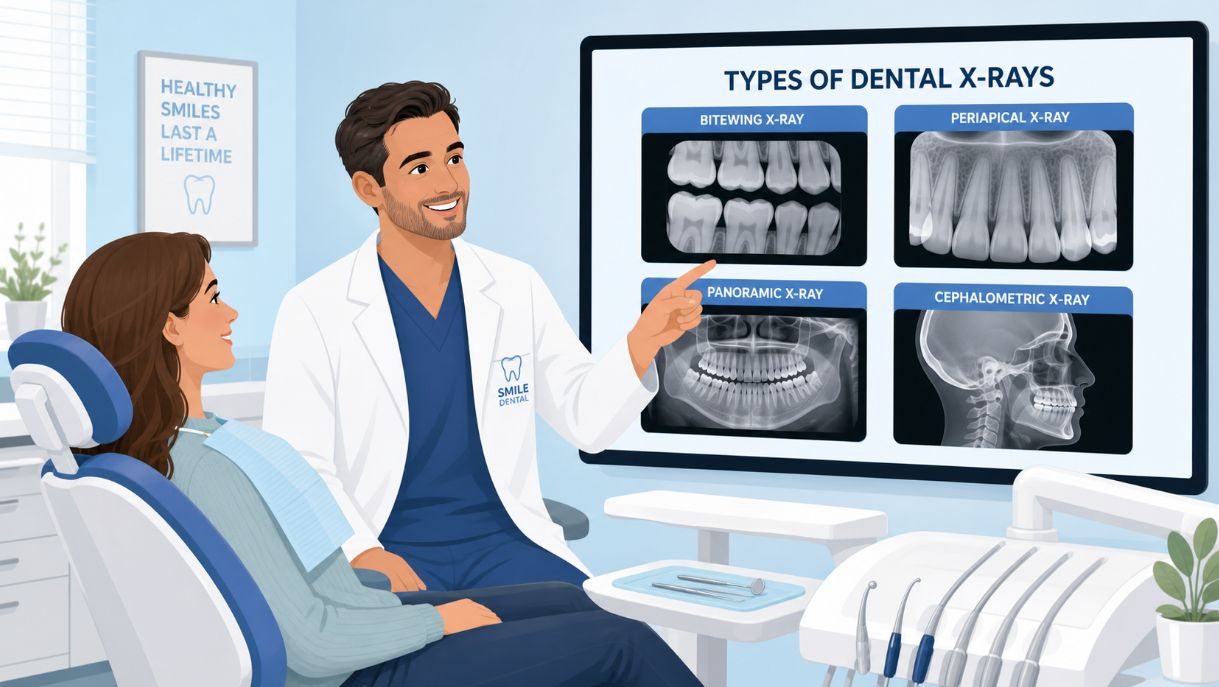
Bitewing (interproximal) radiographs
Bitewing or interproximal X‑rays show the crowns of the upper and lower teeth together, usually in the back of the mouth on one side. You gently bite on a small tab or “wing” to hold the sensor in place while the image is taken.
Dentists use bitewings to:
-
Detect cavities between teeth that are not visible in the mirror
-
Check for decay under or around existing fillings and crowns
-
Assess bone levels around teeth to monitor gum disease
These are often taken at routine check‑ups on a schedule tailored to your risk of decay and gum disease.
Periapical radiographs
Periapical X‑rays capture the entire tooth from crown to tip of the root and the surrounding bone. Each image usually shows one or two teeth in detail.
They are particularly useful to:
-
Investigate severe toothache or suspected abscess at the root
-
Look for cracks, unusual root shapes or extra roots
-
Monitor teeth that have had root canal treatment
-
Evaluate bone around teeth in areas of localised gum disease
Periapical radiographs also form part of a full‑mouth survey (FMX) when your dentist needs a complete baseline record of every tooth.
Occlusal radiographs
Occlusal X‑rays are larger images that show almost the entire arch of teeth in the upper or lower jaw, along with the surrounding bone. You bite gently on a larger film or sensor while the X‑ray beam passes from above or below.
Dentists and especially paediatric dentists use occlusal views to:
-
Assess tooth development in children, including teeth that have not yet erupted
-
Locate extra teeth, impacted teeth or retained roots
-
Examine the floor of the mouth or the palate for cysts, stones or other pathology
These images are less common at routine adult check‑ups but can be invaluable in specific situations.
Extraoral radiography: the bigger picture
Panoramic radiographs (OPG)
A panoramic X‑ray (often called an OPG) provides a wide, single image of the entire upper and lower jaws, all teeth, and the surrounding structures. You stand or sit in the machine while it rotates gently around your head.
Panoramic radiographs are used to:
-
Assess the position and development of wisdom teeth
-
Evaluate the general pattern of all teeth, including impacted or missing teeth
-
Plan extractions, orthodontic treatment and some types of surgery
-
Screen the jawbone and sinuses for cysts, tumours or other abnormalities
They do not show fine detail as clearly as intraoral films, but they offer an excellent overview that guides further imaging if needed.
Cephalometric radiographs
Cephalometric radiographs, or “cephs”, are side‑view X‑rays of the head, showing the teeth, jaws and facial profile together. These are mainly used by orthodontists when planning braces or other bite corrections.
Cephalometric images allow specialists to:
-
Measure jaw growth and relationship between upper and lower jaws
-
Assess how teeth sit in the jaws relative to the rest of the face
-
Plan how orthodontic treatment will move teeth and affect the profile
They are a key part of comprehensive orthodontic assessment in both children and adults.
3D and advanced dental imaging
Cone beam computed tomography (CBCT)
Cone beam CT has transformed many areas of modern dentistry by providing three‑dimensional images of the teeth, jawbones and related structures at relatively low radiation doses compared with conventional medical CT.
CBCT scans are typically used to:
-
Plan dental implants by assessing bone volume, density and exact nerve positions
-
Evaluate complex root canal anatomy or suspected root fractures
-
Investigate impacted teeth and their relationship to nerves and neighbouring teeth
-
Assess jaw joints (TMJs) or trauma where standard X‑rays are not sufficient
Because they carry a higher dose than standard 2D views, CBCT scans are reserved for cases where 3D information will clearly change management.
Conventional CT, MRI and other modalities
In some maxillofacial and hospital dental settings, conventional CT, MRI or other imaging such as ultrasound or PET may be used for complex disease, tumours or jaw joint disorders. These are less common in routine high‑street practice but play an important role in specialist care.
Digital vs traditional film radiography
Most UK practices now use digital sensors instead of traditional film for dental X‑rays. Digital radiography offers several advantages:
-
Lower radiation doses in many cases
-
Instant images on screen, reducing waiting time
-
Ability to enhance contrast and zoom in to detect subtle changes
-
Easy, secure electronic storage and comparison over time
Despite these benefits, the principles of justification and dose limitation are the same, and dentists still follow national guidance to avoid unnecessary exposures.
Safety, radiation dose and UK guidelines
Understandably, many patients worry about radiation. In dentistry, doses are small and tightly controlled. Government reports and professional guidelines track patient doses and equipment trends, showing that typical dental exposures remain low and continue to fall as technology improves.
Key safety principles include:
-
Only taking radiographs when they will genuinely help diagnosis or treatment
-
Using lead shielding and well‑collimated beams where appropriate
-
Choosing the smallest field of view and lowest settings that produce a diagnostically useful image
-
Tailoring frequency of X‑rays to each patient’s risk level, not using a “one size fits all” schedule
For most patients, the benefit of detecting disease early and planning accurate treatment far outweighs the tiny risk from dental X‑ray doses.
How dentists choose the right type of radiograph
Dentists do not simply order “an X‑ray” — they select the most appropriate type based on your symptoms, dental history and overall risk.
For example:
-
Routine check‑up in a low‑risk adult:
-
Bitewing radiographs at intervals based on decay risk to monitor hidden cavities and bone levels.
-
-
Localised toothache:
-
A periapical image of the painful tooth and surrounding area to look for infection, cracks or bone changes.
-
-
Planning braces for a teenager:
-
A panoramic image to see all teeth and roots, plus a cephalometric radiograph to analyse jaw relationships.
-
-
Considering dental implants:
-
Panoramic and often CBCT imaging to measure bone and map nerve positions accurately.
-
UK guidance on selection criteria helps clinicians balance the diagnostic value of each type of radiograph against its dose, so you receive focused, personalised imaging rather than routine exposure.
What to expect as a patient
Modern dental imaging is quick and comfortable. Intraoral X‑rays may involve holding a small sensor between your teeth for a few seconds, while extraoral images like panoramic X‑rays or CBCT require you to stand still while the machine moves around your head.
You will usually be asked to remove jewellery or glasses and to stay as still as possible so the image is sharp. Results appear almost instantly on screen in digital systems, allowing your dentist to show you what they see and explain any findings in real time.
Why understanding radiographs helps you take control of your oral health
When you know what each type of dental radiograph is for, it becomes easier to understand your dentist’s recommendations and to ask the right questions.
You might ask:
-
“What are you looking for with this X‑ray?”
-
“Is this an intraoral or extraoral image, and why did you choose it?”
-
“How often will I need this type of X‑ray based on my risk?”
-
“Can you show me what you see on the radiograph?”
Clear explanations backed by high‑quality images help you stay engaged with your care and make confident decisions about treatments such as fillings, root canal therapy, extractions, implants or orthodontics.